01
Mutate systematically
Selected residues are replaced by alanine to reduce side-chain complexity.

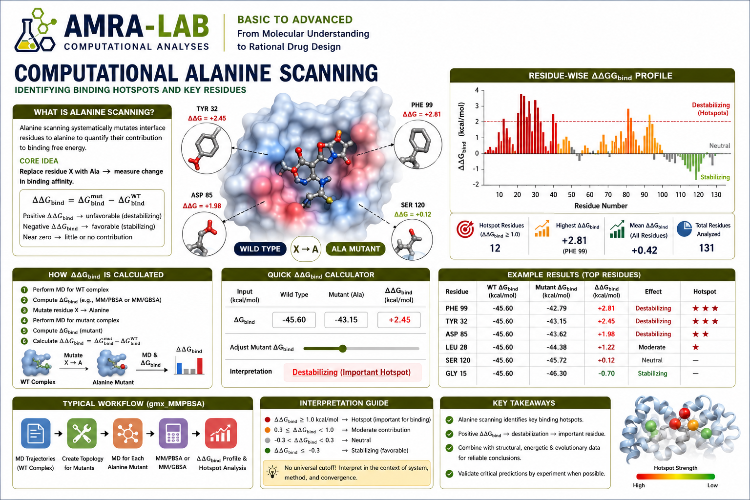
A complete guide to residue hotspot identification, mutation-induced binding-energy changes, ΔΔG interpretation, per-residue decomposition, and responsible reporting after molecular dynamics simulations.
Selected residues are replaced by alanine to reduce side-chain complexity.
Wild-type and mutant binding energies are compared.
Large unfavorable changes after mutation suggest important binding residues.
It is an in silico mutational analysis used to estimate how individual amino-acid side chains contribute to molecular binding or structural stabilization.
A selected residue is computationally changed to alanine. Because alanine has a small methyl side chain, most side-chain-specific interactions are removed while the protein backbone remains largely unchanged.
The change in calculated binding free energy between the wild-type and alanine mutant is reported as ΔΔG.
Identifies residues whose side chains contribute strongly to ligand or protein binding.
Helps select residues for experimental mutagenesis and validation.
Reveals residues that stabilize a ligand and can guide lead optimization.
Estimate ΔGbind for the original complex using the chosen MM/PBSA, MM/GBSA, or another free-energy method.
Replace the selected residue side chain with alanine while preserving the backbone and appropriate topology.
Estimate ΔGbind for the mutant using the same frames, parameters, dielectric settings, and protocol.
ΔΔGbind = ΔGbind,mutant − ΔGbind,wild type.
Residues are ranked by the magnitude and sign of ΔΔG.
Adjust hotspot strength to see how ΔΔG values change.
Use the same analysis protocol for both states.
| Quantity | Value |
|---|---|
| Wild-type binding energy | −42.0 |
| Mutant binding energy | −35.0 |
| ΔΔGbind | +7.0 kcal/mol |
| Interpretation | Likely hotspot |
| ΔΔG pattern | Typical meaning | Caution | বাংলায় |
|---|---|---|---|
| Large positive | Mutation weakens binding; residue may be a hotspot. | Threshold depends on method and uncertainty. | Binding দুর্বল হয়েছে; residue গুরুত্বপূর্ণ হতে পারে। |
| Small positive | Modest favorable contribution in wild type. | May fall within computational noise. | সামান্য contribution। |
| Near zero | Little predicted effect. | Compensating terms may hide interactions. | Mutation-এর effect খুব কম। |
| Negative | Alanine mutant appears more favorable. | Could indicate steric relief, model limitations, or reorganization. | Mutation binding improve করেছে বলে estimate হতে পারে। |
&general
  startframe=1, endframe=500, interval=5,
/
&gb
  igb=5,
/
&alanine_scanning
  mutant_res='A:45,A:78,A:102',
  mutant='ALA',
/
Exact syntax and options depend on the installed gmx_MMPBSA version. Always check the matching documentation and validate residue numbering.
It is a model-dependent estimate and should be interpreted with uncertainty and validation.
Different frames, dielectric constants, or atom selections make the comparison unreliable.
A mutation may cause local rearrangement that a single-trajectory approximation cannot fully capture.
Small positive values may be within noise. Use confidence intervals or replicate/block analysis.
These residues have special backbone roles; alanine substitution may alter conformational behavior beyond side-chain removal.
Special chemistry can invalidate a simple alanine mutation model.
Computational alanine scanning estimates how much a residue side chain contributes to binding under a defined energy model. Large positive ΔΔG values may indicate hotspots, but conclusions should be supported by structural interactions, replicate sampling, uncertainty analysis, and experimental evidence where possible.
Educational content for AMRA-LAB Computational Analyses · Alanine Scanning module