01
Identify donor and acceptor
Find a donor atom bonded to hydrogen and a compatible acceptor atom with available electron density.
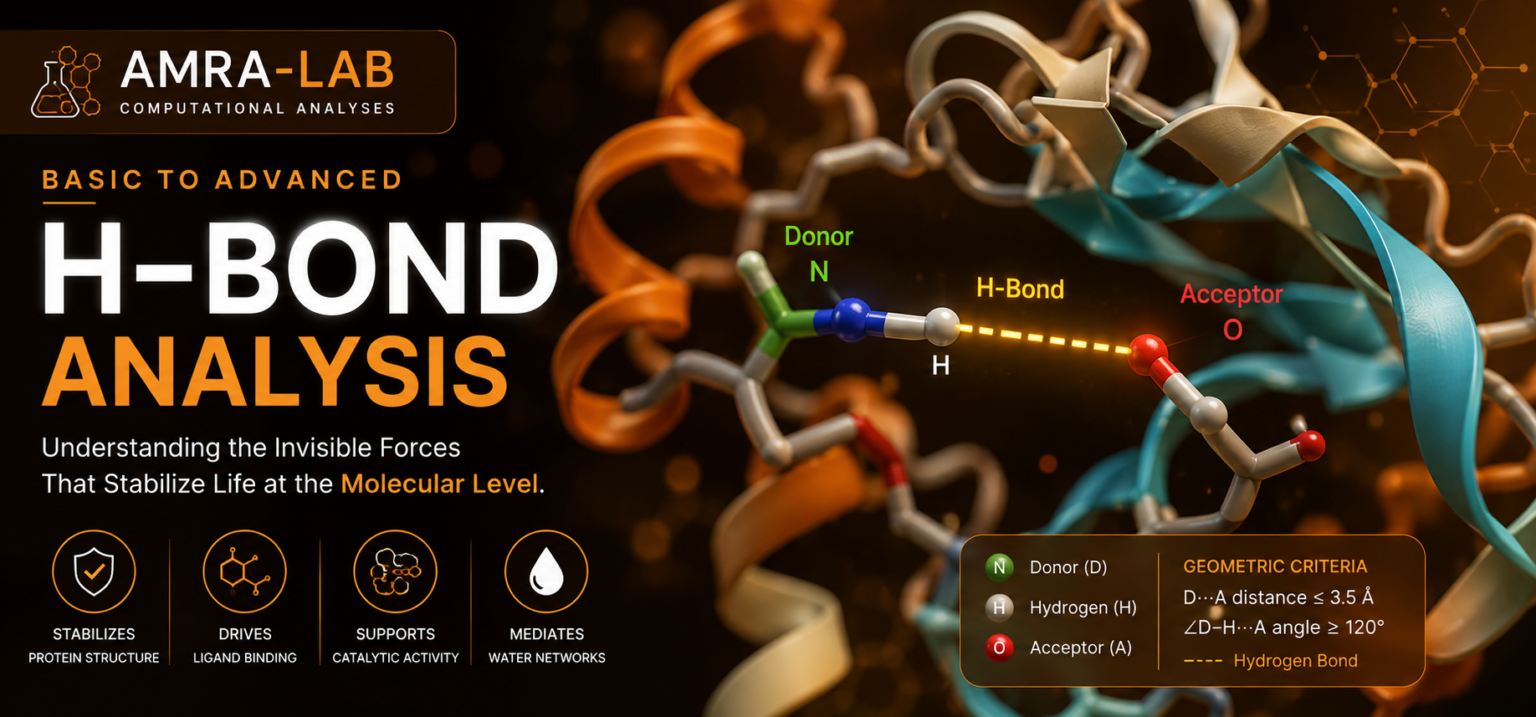
What hydrogen bonds are, how geometrical criteria detect them, why they matter in proteins and protein–ligand complexes, how to interpret their number, occupancy, distance, angle, persistence, and when H-bond analysis can mislead you.
Find a donor atom bonded to hydrogen and a compatible acceptor atom with available electron density.
Measure donor–acceptor distance and hydrogen-bond angle for every trajectory frame.
Study counts, occupancy, distributions, lifetimes, residue identity, and structural context together.
A hydrogen bond is a directional non-covalent interaction involving a hydrogen donor and an electronegative acceptor.
A typical hydrogen bond is written as D–H···A. The donor atom D is covalently bonded to hydrogen H, while the acceptor A provides electron density that interacts with the polarized hydrogen.
In proteins and biomolecular simulations, common donors include O–H and N–H groups. Common acceptors include suitable oxygen and nitrogen atoms, but the exact classification depends on protonation state, chemical environment, force-field topology, and software rules.
Backbone hydrogen bonds support helices, sheets, turns, and many local structural motifs.
Direct and water-mediated H-bonds can help orient a ligand and contribute to binding specificity.
Water can compete with, replace, or bridge protein–ligand and intraprotein hydrogen bonds.
Trajectory software usually applies a geometric rule to each possible donor–hydrogen–acceptor triplet.
Select chemically valid donor atoms that carry bonded hydrogens.
Select atoms capable of accepting a hydrogen bond according to chemical and software definitions.
Calculate the distance between D and A. If it exceeds the chosen cutoff, that frame is not counted as bonded.
Evaluate the D–H···A geometry. More linear arrangements generally represent better directional alignment.
Store 1 when the triplet satisfies all selected criteria and 0 when it does not.
Calculate total count, occupancy, donor–acceptor distribution, angle distribution, persistence, and pair identity.
Move the donor–acceptor distance and angle to see when a candidate interaction satisfies the selected geometric rule.
The bond turns green only when both criteria are satisfied.
Every frame contributes either bonded = 1 or not bonded = 0.
Example: a bond present in 750 of 1000 frames has 75% occupancy.
This interaction appears in most analyzed frames, but its biological importance still depends on residue identity, geometry, alternatives, water competition, and replicate consistency.
Measure H-bonds within the protein to study secondary-structure support, domain contacts, loop closure, salt-bridge-associated polar networks, and mutation-induced rearrangement.
Measure direct contacts between protein donors/acceptors and ligand acceptors/donors. Report pair identity and occupancy, not only the total number.
Useful for solvation, exposed polar groups, desolvation, hydration changes, and competition between water and direct binding contacts.
A bridging water simultaneously connects two partners through two hydrogen bonds. Direct pair analysis alone does not automatically identify complete bridge networks.
These are central to helices and beta sheets. Combine with secondary-structure analysis because simple counts do not uniquely define structural motifs.
Analyze active-site residues, proton relays, catalytic triads, conserved waters, and residues controlling substrate positioning.
The total number fluctuates around a similar range. This suggests a broadly persistent network, but individual bonds may still exchange rapidly.
May indicate structural expansion, loss of contacts, solvent competition, ligand movement, or a conformational transition. It is not automatically evidence of instability.
May reflect compaction, formation of new polar contacts, water-network reorganization, or ligand adaptation. More bonds are not always better.
Frequent formation and breakage may indicate flexible contacts, alternate donor–acceptor partners, solvent exchange, or multiple conformational states.
| Metric | Meaning | Useful for | Major limitation |
|---|---|---|---|
| Instantaneous count | Number of H-bonds in each frame. | Overall time evolution. | Does not identify which bonds are present. |
| Pair occupancy | Percentage of frames satisfying the criterion. | Persistent residue or ligand contacts. | Depends on cutoff, sampling, and intermittent rebinding. |
| Continuous lifetime | Duration before a bond first breaks. | Uninterrupted persistence. | Very sensitive to brief geometric interruptions. |
| Intermittent lifetime | Allows temporary breaking and rebinding under a defined model. | Dynamic contact memory. | Requires explicit methodological definition. |
| Distance distribution | Distribution of donor–acceptor separation. | Geometry quality and alternate states. | Distance alone is insufficient. |
| Angle distribution | Distribution of directional alignment. | Bond geometry and orientation. | Angle convention varies among tools. |
Incorrect. Binding depends on desolvation, geometry, competing interactions, entropy, electrostatics, conformational strain, and the surrounding environment.
A wrong protonation or tautomer assignment changes donor and acceptor identity and may create or remove apparent hydrogen bonds.
Average count hides pair exchange. Report important donor–acceptor identities, occupancy, distributions, and representative structures.
For GROMACS group-based analysis, selected groups must meet the tool requirements; identical or non-overlapping groups are expected.
Some programs use deviation from linearity; others report the D–H···A angle directly. A cutoff of 30° in one convention may correspond to 150° in another representation.
Broken molecules or complexes across the box can generate false distances or missed interactions. Process and visually inspect the trajectory.
A catalytic H-bond, a surface water contact, and a transient terminal contact do not have equal structural significance.
Commands vary between the current selection-based tool and the legacy implementation. Confirm the syntax supported by your installed GROMACS version.
gmx trjconv -s md.tpr -f md.xtc -o md_whole.xtc -pbc mol -center
Choose a suitable centring group and output group. Verify membrane proteins and multimers visually because generic processing may be inappropriate.
gmx hbond -s md.tpr -f md_whole.xtc -r 'group "Protein"' -t 'group "Protein"' -num hbonds_protein.xvg -dist hbdist_protein.xvg -ang hbang_protein.xvg
The current tool supports reference and target selections. The selections must be identical or non-overlapping.
gmx hbond -s md.tpr -f md_whole.xtc -r 'group "Protein"' -t 'resname LIG' -num hbonds_protein_ligand.xvg -dist hbdist_protein_ligand.xvg -ang hbang_protein_ligand.xvg
Replace LIG with the actual ligand residue name. Validate donor/acceptor assignment from the topology and chemistry.
gmx hbond -s md.tpr -f md_whole.xtc -r 'group "Protein"' -t 'resname LIG' -hbr 0.35 -hba 30 -num hbonds_PL.xvg
Here -hbr controls the H-bond distance cutoff and -hba controls the angular cutoff in the current GROMACS definition.
gmx hbond-legacy -s md.tpr -f md_whole.xtc -n index.ndx -num hbnum.xvg -dist hbdist.xvg -ang hbang.xvg
Legacy output can include autocorrelation and lifetime-related analyses. Do not mix current and legacy syntax without checking your version.
gmx hbond -h and record your software version before analysis.“Protein–ligand hydrogen bonds were analyzed over the production trajectory after periodic-boundary correction using a donor–acceptor distance cutoff of 0.35 nm and the stated angular criterion. The number of hydrogen bonds, donor–acceptor distance distributions, angle distributions, and pair occupancies were evaluated.”
No. It means the chosen geometry is frequently satisfied. Importance depends on location, chemistry, alternatives, desolvation, function, and reproducibility.
Depending on chemistry and geometry, a donor or acceptor can participate in alternative or simultaneous interactions. Interpret unrealistic over-coordination carefully.
Hydrogen bonds are dynamic and geometric cutoffs convert continuous motion into a binary state. Small movements near a cutoff can cause frequent switching.
Include water when hydration, competition, or bridging matters. Exclude it when you specifically want direct protein–ligand contacts, but state that choice.
No. A ligand may maintain H-bonds while drifting, or remain bound through hydrophobic and ionic interactions with few hydrogen bonds.
They may use different donor/acceptor rules, atom naming, distance definitions, angle conventions, cutoffs, protonation states, and PBC handling.
Not directly. Larger systems have more possible donors and acceptors. Normalize or focus on equivalent regions and matched atom selections.
Hydrogen-bond analysis describes how often chemically and geometrically defined donor–hydrogen–acceptor arrangements occur. It does not independently measure binding free energy, global stability, or biological activity.
Educational content for AMRA-LAB Computational Analyses · Hydrogen-Bond Analysis module